- Over 55,000 LASIK and cataract procedures (including on over 4,000 doctors)
- The FIRST center in TN to offer Laser Cataract Surgery
- Introduced bladeless all-laser LASIK to the state
- Implanted the state's first FOREVER YOUNG™ Lens
- The first surgeons in the US to perform a new Intacs surgery to treat keratoconus
- Helped patients from 40 states and 55 countries
- International referral center for cataract surgery and LASIK complications
- Read Dr. Wang's book: LASIK Vision Correction
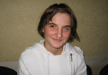
Why did you decide to have LASIK? Why did you choose Dr. Wang? How has your life changed since your LASIK procedure?
What is your advice for people considering LASIK?
Click to read more


Chapter ElevenOther Refractive ProceduresWhile the most popular and widely practiced vision correction procedure performed today is LASIK, surgeons have access to a wide variety of refractive procedures that may be employed, should your particular needs not fall into the realm of LASIK. Here are some additional refractive procedures and what is involved in each of them, starting with photorefractive keratectomy (PRK), the second most widely practiced laser vision correction procedure performed today. Photorefractive Keratectomy (PRK) What to Expect PRK is relatively easy for your surgeon to perform. The real work for the doctor and patient begins after the procedure when the doctor has to very carefully monitor the healing process. Laser Vision Correction with PRK The procedure for laser vision correction with PRK is very similar to that of LASIK. The biggest difference is that no microkeratome is used to create a corneal flap. Instead, the excimer laser makes its correction directly on the surface of your cornea, removing the corneal epithelium in the process. The actual laser part of the procedure takes twenty to ninety seconds. At the end of the procedure, a clear-bandage contact lens is placed onto your eye to help keep you comfortable while the corneal epithelium regenerates (usually three to five days). A typical PRK procedure takes about five to ten minutes per eye. Operating on one eye or both eyes on the same day is a decision that is made by the patient after discussing the pros and cons of this option with the surgeon. Because the return of functional vision is sometimes prolonged under PRK, most surgeons prefer to wait at least one week before operating on the second eye. Previous Surgery Patients who have previously undergone certain types of surgery are sometimes a candidate for PRK as a second procedure to enhance the visual results. Post Correction Care Once your PRK procedure is completed, you will sit with your eyes closed for thirty minutes. Afterward, the doctor will check your eyes; then you will be instructed to go home and take a nap. Because your vision will be somewhat blurry, you will need to have someone drive you. Additional drops will be placed in your eyes before you go, and you will be instructed on the use of your medications. You may also be given clear plastic shields to wear at night for as long as your doctor instructs. These prevent accidental trauma to your eye while sleeping. Before you go home to rest, your surgeon may also give you something to help you sleep, for sleep is the best way to keep your eyes closed for a few hours. Following the PRK surgery, you may experience some discomfort and blurriness which will last for a few days. Tylenol®, aspirin, ibuprofen, or similar over-the-counter pain medications are usually adequate to keep patients comfortable. Your doctor may give you a prescription for a stronger pain medication, such as Vicodan®, in case your pain is more severe, and something to help you sleep. You will also be given antibiotic eye drops, anti- inflammatory eye drops, and lubricant eye drops to promote healing. Nonpreserved anesthetic drops sometimes may be used sparingly, no more than four or five times over the first twenty-four hours. Excessive use of anesthetic drops is toxic to the cornea and will prevent healing. You can expect your vision to be fairly blurry, and your vision may worsen over the first three days. It will then begin to improve. Guidelines for Healing Follow these guidelines to promote safe and rapid healing:
Postoperative Follow-up Schedule When you leave the laser center, you will be given complete instructions to follow, including a postoperative appointment schedule similar to the one shown on the next page. |
| Purpose of Appointment | Time Frame Following PRK Procedure |
|
1 day |
|
3-4 days |
|
1 week - 1 month |
|
2-3 months |
|
4, 6, 12 months |
|
The PRK Recovery Cycle Your vision may get worse for the first three days. However, you will notice an improvement in your vision by about day five, and it will continue to improve for several weeks. Most patients can return to work and resume most normal activities after three to five days. However, it may take as long as three months for your vision to stabilize. During this period, you must continue to use the postoperative steroid drops as your doctor suggests. Deviations from this regimen may lead to haze (scarring of the cornea) or regression of the refractive result. You may be asked to use the drops for up to three months; the exact frequency of usage will be modified at the one-month visit. Be sure to keep your follow-up appointments as your drop regimen may be altered. Also, the doctor needs to monitor your eye pressure while you are on postoperative steroids. With PRK, your vision usually becomes completely stable within six to twelve months. Once your vision is stable, your treatment is permanent. You now have less dependence on, and maybe complete freedom from, glasses and contact lenses. Complications with PRK The potential complications that may arise from PRK are similar to those in LASIK (see Chapter Ten, "Risks and Complications"). They include unrealistic expectations, undercorrection, overcorrection, induced astigmatism, dry eye, haze, night glare and halos, loss of best corrected vision, regression, and infection or severe inflammation. Regression occurs when a patient appears to be adequately treated on the first few postoperative visits, but over the next several weeks to months begins to return toward the original prescription. The amount of regression is usually small; however, occasionally it is visually significant and requires an enhancement procedure. The enhancement procedure is usually performed three to six months following the original procedure. This allows for the refraction to stabilize. Regression occurs more commonly after PRK than after LASIK, and it is more common after PRK and LASIK in patients with higher amounts of myopia, hyperopia, or astigmatism. Enhancement following PRK involves a prolonged healing cycle similar to the initial procedure. Probably the most common complication following PRK is trace amounts of haze, but even a moderate amount of haze will not affect your vision. If haze does develop during the postoperative period, your surgeon may increase the frequency of steroid drops or even surgically irrigate your eye to alleviate or lessen the haze. Serious haze occurs in less than one percent of all patients who have undergone PRK; it is more common with higher corrections but can occur with low corrections. Symptoms of serious haze can by eliminated with treatments that mechanically remove the haze concurrent with applications of the drug Mitomycin C. Pros and Cons of PRK Pros
Cons
IntacsTM Corneal Ring Segments Approved in April 1998, the Intacs corneal ring segments offer patients with mild myopia and minimal astigmatism another option for correcting their nearsightedness. Currently, the rings are approved for correction of nearsightedness up to 3.00 diopters and astigmatism up to 1.00 diopter in patients twenty-one years or older who have a stable prescription. The procedure involves placing two small plastic "ring" segments in the peripheral cornea; this allows the central cornea to flatten. The advantage of Intacs is that the tiny segments may be removed if the patient wishes to reverse the correction. In most patients in the clinical trials, when the rings were removed, the eyes went back to their preoperative state. In a few patients, they did not. Because of those few patients whose eyes did not return exactly to their preoperative condition, the FDA will not allow the use of the term "reversible," but Intacs are removable if necessary. Intacs insertion is accomplished in a time frame similar to LASIK's, taking roughly fifteen minutes per eye under numbing drops. The recovery of clear vision seems to take slightly longer than LASIK and doesn't seem to have quite the "wow" effect of rapid visual recovery. The procedure is newer than LASIK or PRK, so it doesn't yet have the track record that the other two procedures do. The cost of Intacs is roughly equal to or more than LASIK in most centers. Removal of the rings, either for fine tuning the result or from dissatisfaction, is accomplished with a second surgery. The segments are removed, the eyes are allowed to heal, and an alternate procedure (such as LASIK or PRK, or a change in ring size) may be performed once the eyes have healed. The treatment range for Intacs is currently very limited, and astigmatism cannot be treated by them; hence, patients with astigmatism are not good candidates for Intacs. Astigmatic Keratotomy (AK) Astigmatic keratotomy (AK) is similar to RK, but its purpose is to correct astigmatism. Incisions are made in the cornea in such a way as to make it more round (analogous to loosening the laces on a football). This procedure is often combined with radial keratotomy (RK) and has a similar long track record. AK is still considered an excellent procedure for correcting pure astigmatism (patients without coexisting nearsightedness or farsightedness). AK can also be used to enhance the results of LASIK and PRK by correcting small residual amounts of astigmatism. However, most surgeons prefer to correct astigmatism with the excimer laser. Cataract Surgery For patients with significant cataracts who are looking for an option for correcting their nearsightedness or farsightedness, cataract surgery presents the best option for achieving this goal. After removing the cataract with ultrasonic power, the surgeon can implant a lens that will reduce or eliminate nearsightedness or farsightedness. This procedure is not performed in younger patients without cataracts because the surgery involves entering the eye and, therefore, slightly increases the risk of more serious complications. The surgery also involves removing the natural crystalline lens, which in young people allows them to focus up close. LASIK surgery, which leaves the lens intact, is a better option for younger patients. Automated Lamellar Keratoplasty (ALK) Automated lamellar keratoplasty (ALK) is a refractive procedure that was done on high myopes prior to the invention of the excimer laser. It is similar to LASIK in that it uses a special instrument called a microkeratome to separate the surface layer of the cornea. This "flap" is temporarily folded back (similar to the first part of the LASIK procedure), and a thin disc of corneal tissue is removed with a second pass of the microkeratome. The procedure is much less precise than LASIK and is associated with a much higher complication rate. It is primarily used to correct large amounts of myopia. Satisfactory results are not always obtained the first time through, and a high percentage of eyes needs additional procedures to achieve the desired result. Sometimes an irregular corneal surface results from the procedure, causing some distortion of vision. Automated lamellar keratoplasty is rarely performed today because of the advent of LASIK. LASIK has essentially replaced ALK because of the increased accuracy afforded by the excimer laser in making the second "cut." Radial Keratotomy (RK) Until recently, radial keratotomy (RK) was the most commonly performed refractive procedure for nearsighted patients. With the aid of a high-powered microscope, the surgeon makes a series of radial microscopic incisions (usually between four and eight) on the surface of the cornea in order to reduce its curvature. This procedure is well suited for patients with low myopia and has been used for over twenty-five years. Although rarely performed anymore because of the increased accuracy and stability of the excimer laser techniques, RK is still an effective vision correction technique and is used in those areas of the world that do not have access to the much more expensive laser technologies. Bioptics Bioptics is a combination procedure involving a phakic intraocular lens implant followed by LASIK. It is recommended for the most extreme levels of myopia and hyperopia when neither technique alone will entirely correct the refractive error. This combined technique can be used to correct over 30.00 diopters of myopia--nearly three times the maximum amount of myopia that can be safely corrected with LASIK. Clear Lens Extraction (CLE) Clear lens extraction (CLE) involves removing the internal lens of the eye, just like in a cataract operation. This is done with a special ultrasound instrument and may be done with eye-drop anesthesia (similar to LASIK). The procedure can be performed without the need for stitches. A flexible synthetic lens implant of the proper power is then placed inside the eye through an extremely small incision to correct the refractive error. This procedure more commonly is performed for treating higher levels of farsightedness in patients over age forty. The optical results are excellent and the visual recovery period is brief. Clear lens extraction may also be used to correct higher levels of nearsightedness and may be fine tuned with LASIK if a small refractive error remains. Some surgeons have used this procedure to treat extremely nearsighted or farsighted patients who are not candidates for PRK or LASIK. The major drawbacks of this procedure are the increased risk of postoperative retinal detachment, the increased risk of intraocular surgery, and that patients usually need reading glasses afterward. A new intraocular lens called the ARRAY is a multifocal lens that can be implanted at the time of lens extraction. The ARRAY intraocular lens allows you to see both near and far after the operation. In order for it to work to its maximum potential, both eyes should be implanted with the lens. Because of its multifocal capacity, some patients experience a loss of contrast at night and also develop halos around lights. If these symptoms become problematic, the ARRAY lens can be removed and replaced. Conductive Keratoplasty (CK) Conductive keratoplasty utilizes a special probe that introduces an electrical current to the peripheral cornea, inducing a tightening of the corneal fibers. This acts like tightening a belt, causing the central cornea to steepen. It is effective for small amounts of hyperopia. A few ophthalmologists are currently conducting the FDA clinical trial of this procedure and have been impressed with its safety and effectiveness. Laser Thermal Keratoplasty (LTK) For low amounts of farsightedness, a technique called laser thermal keratoplasty (LTK) is a possible method of thermally changing the shape of the cornea. A special holmium laser is used to place laser "burns" in the peripheral cornea to slightly tighten the fibers and thereby steepen its curvature. The technique only seems to work for low amounts of farsightedness and recently received FDA approval in January 2000. Although not a treatment for presbyopia, some physicians are using LTK to create monovision in patients, thus relieving the need for reading glasses. Phakic Intraocular Lens (PIOL) Implants Implantable contact lens technology has arisen out of the incredible advances in modern cataract surgery. Current technology allows ophthalmologists to insert flexible intraocular lenses (used to replace the natural lens after cataract surgery) through extremely small incisions. The lenses are flexible enough to allow folding and insertion through a small incision opening. Once in the eye, the lens expands to its full size, allowing the eye to remain relatively untraumatized, thus reducing astigmatism and recovery time. This same "small incision" lens technology allows the surgeon to insert an even thinner, foldable lens in front of the natural lens to correct nearsightedness or farsightedness. A phakic intraocular lens (PIOL) is a lens implanted inside the eye for the correction of either extreme nearsightedness or extreme farsightedness. In effect, the lens becomes an internal contact lens rather than a contact lens on the surface of the eye. It is usually recommended for patients whose visual correction is outside the range that can safely be treated with LASIK. Because of the slightly increased risk of more serious complications, the PIOL is reserved for high amounts of nearsightedness or farsightedness--above the current limits of LASIK. In places where this technology is available, surgeons are implanting the PIOL in patients with myopia greater that 12.00 to 15.00 diopters and hyperopia greater than 4.00 to 6.00 diopters. Despite the excellent outcomes in most cases, complications associated with the implants are currently the biggest concern. Specifically, in the early studies a small percentage of patients developed cataracts shortly after implantation of the lens. There is also a small risk of endophthalmitis (infection within the eye) because the surgical incision actually enters the eye. This complication can lead to complete loss of vision. Endothelial cell loss with some lens designs is also a concern and is being studied rigorously. Some ophthalmologists in the United States are currently performing this procedure as part of an FDA clinical trial. The procedure holds a lot of promise for extremely nearsighted or farsighted individuals. Ophthalmologists are anxious to see how the implantable lenses fare in current studies using newer lens designs and implantation techniques. These lenses are currently being used in Europe and South America with very high success. The results will be presented to the Food and Drug Administration with the hope that the FDA will authorize other eye surgeons to use this exciting new technology. Surgery for Presbyopia One of the more exciting areas of ophthalmology is the surgical treatment of presbyopia, the stiffening of our natural lens that decreases near vision as we age. Several devices and surgeries have been tried, all of which direct their effect at enlarging the circumference of the front of the eye and tightening the fibers that control the focus of the lens. One of the principle theories of presbyopia suggests that as we age these fibers stretch, becoming less effective. By enlarging the circumference of the eye, the fibers should once again become tight and thus effective at focusing the lens. Anterior Ciliary Sclerotomy (ACS) Anterior ciliary sclerotomy is a surgical procedure for relieving presbyopia. It involves the creation of several small incisions in the sclera (coating of the eye) directly over the muscle that controls the eye's natural lens. The purpose of this procedure is to expand the circumference around the equator of the eye. Scleral Expansion Bands (SEB) Scleral expansion bands (SEB) is another procedure for relieving presbyopia. It consists of a number of thin silicon bands implanted in the sclera to stretch or expand the equator of the eye in order to restore accommodation (the ability to read without corrective lenses after age forty-two). The theory behind both procedures is that expansion of the eye will allow increased room for the eye's natural crystalline lens to move normally, enabling the eye to see near objects again. These procedures are still being investigated in the United States. To date, there is much controversy about both the theory and the effectiveness of these types of surgery. Until scientific studies show more consistent results, monovision and reading glasses or bifocals are still the best options for treating presbyopia. |
If you're interested in bladelessLASIK, choose experience and technology. Choose Wang Vision 3D Cataract and LASIK Center of Nashville, Tennessee. Call (615)321-8881 or email us today.

 |
 |
 |
 |
 |
 |
 |
 |
 |
 |
Our new texbooks

A 501c(3) charity that has helped patients from over 40 states in the US and 55 countries, with all sight restoration surgeries performed free-of-charge.