- Over 55,000 LASIK and cataract procedures (including on over 4,000 doctors)
- The FIRST center in TN to offer Laser Cataract Surgery
- Introduced bladeless all-laser LASIK to the state
- Implanted the state's first FOREVER YOUNG™ Lens
- The first surgeons in the US to perform a new Intacs surgery to treat keratoconus
- Helped patients from 40 states and 55 countries
- International referral center for cataract surgery and LASIK complications
- Read Dr. Wang's book: LASIK Vision Correction
Why did you decide to have LASIK? Why did you choose Dr. Wang? How has your life changed since your LASIK procedure?
What is your advice for people considering LASIK?
Click to read more


| Home | Print This Page |
History of LASIK Surgery
in Nashville, Tennessee
Dr. Wang, Refractive Surgeon in Nashville, Tennessee
Forerunners to Modern Refractive Surgery:
The study of visual problems (refractive errors) began in the early sixteenth century when Leonardo da Vinci contemplated the possible source of visual disturbances. But real progress in the field of vision correction was constrained until a better understanding of how the eye functions was acquired.
Several decades later came the advent of topical (eye drop) anesthesia, which led to cataract surgery after the Civil War. In 1867, with the development of the keratometer (an instrument for measuring the curvature of the cornea), surgeons could measure astigmatism following cataract surgery.
In 1869, Snellen, after whom the vision charts of today are named, proposed using incisions across the steep meridian of the cornea to flatten it and treat astigmatism. However, twenty-one years would pass before anyone (Galezowski) would actually attempt, albeit unsuccessfully, to flatten the corneal contour.
Trials and Experimentation:
Not long after a successful cataract surgery technique was developed by Van Graefe in the 1850s, ophthalmologists everywhere began to recognize the impact of corneal shape on astigmatism.
It was about this time that a Dutch physician, Leendert Jan Lans (working at the time on his doctoral degree), began to systematically study and define the principles of keratotomy. So fundamental and comprehensive was his research that it soon became the standard of refractive surgery. He practiced and promoted the principles of corneal flattening that could be achieved by incisions made on the anterior surface of the cornea. By varying the number, direction and shape of the incisions, Lans could manipulate the effects and tailor the visual correction.
In addition to surgical techniques, there were nonsurgical attempts at reducing myopia by manipulating the shape of the eye. One remedy was an eyecup with a spring-powered mallet designed to flatten the cornea; another was a firm rubber band used to flatten it. But these were techniques that failed to result in any significant degree of visual correction.
With the exception of the work performed by Lans, 1885 to 1939 was principally a time of trial and error for refractive surgery. Nevertheless, the successes and failures of this period helped determine which refractive procedures worked and which did not.
Modern Refractive Surgery:
In 1936, Tsutomu Sato observed a flattening of the cornea in patients who had sustained traumatic injury to the eyes. His work led numerous assistants to establish the value of radial keratotomy, built upon the principles outlined by Lans nearly half a century earlier and applied to the treatment of keratoconus corneas. Sato brought anterior and posterior keratotomy to clinical practice in hundreds of patients and reported his results in the early 1940s. Other ophthalmic surgeons subsequently used his technique and obtained similar results.
In 1948, Ridley, a physician to Royal Air Force pilots in World War II, noted that pilots whose eyes harbored slivers of Perspex (cockpit "glass") seemed to have little or no reaction to this foreign material. This led him to suppose that a small lens made out of the same material could probably be tolerated inside the human eye. Soon he began experimenting with plastic lens designs, and the modern era of intraocular lens implantation for cataract surgery was born.
About the same time that Ridley envisioned the plastic intraocular implant, José Barraquer in Columbia developed the idea of lamellar (pancake and flap-based) corneal surgery to alter the shape of the cornea.
In 1949, Barraquer described the principles of lamellar surgery. He changed the cornea's shape by removing the anterior cornea (the equivalent of today's corneal flap) with an instrument called a microkeratome, freezing it and changing its shape with a mechanical lathe called the cryolathe. In the mid-1980s, the cryolathe rose to its highest state of precision through automation. In 1985, Casimir Swinger developed a method of changing the shape of the cornea without freezing it. He did this using the microkeratome only. Then in 1987, Luis Ruiz, a protégé of Barraquer, modified the principles of microkeratome corneal resection by using an automated form of the instrument to perform the operation directly on the eye. This procedure, called automated lamellar keratoplasty (ALK), was used to correct high levels of myopia and hyperopia.
Halfway around the globe, a handful of Russian ophthalmologists began research to determine whether or not RK (radial keratotomy, or straight-line incisions placed in a spoke-like pattern around the periphery of the cornea) could be effective if it was confined to the anterior side of the cornea. This would thereby avoid the long-term problems that arose from the disruption of the corneal endothelium in Sato's posterior keratotomies.
By the mid-1970s, Russian scientists such as Durney, Yenaleyev and Fyodorov had determined that most of the radial keratotomy flattening effect could be obtained with sixteen or fewer incisions. Fyodorov developed a system of anterior radial keratotomy that, by varying the number of incisions and the amount of uncut clear central zones between them, permitted him to carefully control the degree of visual correction. It was he who convinced the world that radial keratotomy (RK) could indeed reduce or eliminate myopia.
Radial keratotomy was introduced into the United States in 1978 by Leo Bores. It soon became a subject of great interest and careful scientific scrutiny. In 1980, the National Institutes of Health sponsored the PERK (Prospective Evaluation of Radial Keratotomy) study, which provided factual, scientific data on radial keratotomy performed in a standard manner in nine centers across the United States.
The Arrival of the Excimer Laser:
The first step in the evolution of laser surgery occurred when experts researched the application of laser technology to vision correction. In 1980, Beckman and Peyman and their associates used a carbon dioxide laser to create thermal shrinkage of the cornea in order to change corneal contour. A year later in 1981, John Taboada reported at a meeting of the Aerospace Medical Association that the argon-fluoride excimer laser had the ability to indent eye tissue. Work then proceeded on ablation (microsurgical removal) of corneal tissue to flatten the cornea. Further evaluation was performed by Steve Trokel.
The first use of the excimer laser on blind human eyes took place in 1985 by Seiler in Germany. This was followed in 1987 by L'Esperance of the United States. The procedure was called photorefractive keratectomy (PRK) and involved the ablation of the surface of the cornea to flatten its central portion in order to correct nearsightedness. In 1989, Michelson and four other American ophthalmologists traveled to the Free University of Berlin to observe Seiler perform photorefractive keratectomy on patients who were nearsighted. In 1991 Michelson and this elite group of ophthalmologists became the first five clinical investigators of the excimer laser in the United States, utilizing the laser manufactured by Summit Technology.
By 1990 and 1991, Pallikaris and Buratto and their associates had combined lamellar splitting (using the blade of a microkeratome to make a corneal flap, based on Barraquer's pioneering work forty years earlier) with excimer laser ablation of the exposed corneal bed. It was Pallikaris who coined the term LASIK (laser in-situ keratomileusis). This procedure took the moderately successful automated lamellar keratoplasty (ALK) procedure and added the incredible accuracy of the excimer laser to improve the results.
The LASIK procedure avoids the anterior stromal haze and pain generally associated with surface ablation by the excimer laser (PRK). This result is achieved because the laser is applied only within the corneal substance rather than removing a large area of epithelium (the thin, outer surface layer of the eye). When the epithelium is removed during PRK, the nerve endings are exposed; these exposed nerve endings cause pain during recovery. Additionally, there are more fibroblasts underneath the epithelium, and these contribute to scarring. Lastly, the epithelium is the eye's mechanical barrier to bacteria; removing it increases the risk of keratitis (infection). With LASIK, the epithelium remains almost entirely intact. As a result, the nerve endings stay covered and there is minimal pain during recovery. With the epithelium intact and healed within twelve hours after the procedure, there is a lower risk of infection and scarring.
The initial clinical trials of LASIK in the United States began in 1996. These clinical investigations culminated in the approval by the FDA of the LASIK procedure in 1999 in which as a panel consultant for the FDA, Dr. Wang was appointed as a primary reviewer for LASIK for FDA approval.
If you're interested in LASIK, choose experience and technology. Choose Wang Vision 3D Cataract and LASIK Center of Nashville, Tennessee. Call or email us today.

 |
 |
 |
 |
 |
 |
 |
 |
 |
 |
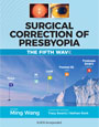
Our new texbooks

A 501c(3) charity that has helped patients from over 40 states in the US and 55 countries, with all sight restoration surgeries performed free-of-charge.