- Over 55,000 LASIK and cataract procedures (including on over 4,000 doctors)
- The FIRST center in TN to offer Laser Cataract Surgery
- Introduced bladeless all-laser LASIK to the state
- Implanted the state's first FOREVER YOUNG™ Lens
- The first surgeons in the US to perform a new Intacs surgery to treat keratoconus
- Helped patients from 40 states and 55 countries
- International referral center for cataract surgery and LASIK complications
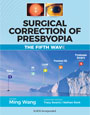
- Read Dr. Wang's book: LASIK Vision Correction
Why did you decide to have LASIK? Why did you choose Dr. Wang? How has your life changed since your LASIK procedure?
What is your advice for people considering LASIK?
Click to read more


| Home | Print This Page |
Discussion: Accommodating IOL patient with residual astigmatism
Case presentation: The patient is a 42-year-old woman who presented in 2008 looking to decrease her dependence on corrective eyewear. At that time, the patient’s ocular and medical history were normal.
Her uncorrected vision was count fingers in each eye. The vision in the right eye was 20/20 with a refraction of –8.25 sphere. The refraction in the left eye was 20/20 with a refraction of –7.75 +0.50 × 065. Her right eye was found to be her dominant eye. Corneal pachymetry was 505 µm in the right eye and 497 µm in the left eye. Slit lamp examination showed no evidence of cataracts and an otherwise normal and healthy anterior segment in both eyes. Fundoscopic examination revealed a normal retinal exam in both eyes.
After an extensive discussion with the patient concerning her high degree of myopia, her asymmetric astigmatism and slightly thin corneas, we compared the options of refractive lens exchange, laser vision correction using surface treatment and no surgery. After careful consideration, the patient underwent successful implantation of bilateral Crystalens HD lenses (Bausch + Lomb) as a refractive lens exchange procedure in each eye in July 2009.
At her 3-month postoperative visit in October 2009, the patient stated her vision was “pretty good for both distance and near.” Uncorrected visual acuity at that time was 20/25+3 in the right and 20/25+2 in the left eye for distance. Uncorrected near vision was J2 in the right eye and J3 in the left eye. The refraction in the right eye was –0.75 +0.75 × 045 yielding 20/20 vision. The refraction in the left eye was –0.75 +0.5 × 065 yielding 20/20 vision. Her distance-corrected near vision with this previous refraction was J5 in the right eye, J3 in the left eye and J3 with both eyes together.
The patient was happy with her results overall but had been noticing a slight weakening in her near visual acuity. She was also interested in trying to sharpen her distance visual acuity even though it was generally doing very well. We discussed at length with the patient the excellent results she had but also described that she had some slight residual refractive astigmatism in the right eye greater than the left eye.
- Would you have counseled the patient differently concerning her procedure choices upon initial presentation? If you would have chosen a refractive lens exchange (RLE) procedure, what type of implant would have been your first choice? If laser vision correction were your choice, what type of procedure would you have chosen?
- Because this patient decided to pursue the Crystalens HD, what next step would you take at her 3-month postoperative visit with her level of visual acuity and current complaints?
Approach 1: Jonathan H. Talamo, MD
RLE for this highly myopic patient with possible subtle forme fruste keratoconus was a reasonable surgical option. Given the corneal topography, refraction and pachymetry measurements, laser vision correction (PRK or LASIK) was clearly not an option, as the already abnormal-appearing corneas would have been left unacceptably thin. The additional surgical option I would have discussed with this patient is phakic IOLs. With an anterior chamber depth greater than 3 mm, a phakic IOL such as the STAAR Visian Implantable Collamer Lens would have been a good option for this patient, as some natural accommodation would have been maintained.
If I had performed RLE for this patient, I would have also recommended a Crystalens IOL. My IOL model of choice would have been the Crystalens 5-0 or AO (not yet available at that time) rather than the HD. The reason for this is that the prolate, asymmetric nature of the corneal topography could potentially interact in a less-predictable-than-normal fashion with the small central add present within the center of the HD IOL optic. I would not have recommended a multifocal IOL for similar reasons.
The visual status in October 2009 clearly indicates that this patient had an excellent response to surgery. I would acknowledge her visual symptoms/complaints but also reassure her that I felt that no further surgery should be done at that time. Furthermore, I would communicate to her that in my opinion her near vision was as good as anyone could expect. I would also encourage her to continue to read without correction and to perform accommodative exercises on a twice-daily basis. Unless slit lamp examination showed capsular contraction syndrome or posterior capsular opacity that I felt was contributing to the patient’s visual symptoms, I would not perform a YAG capsulotomy yet.
To help her better assess whether her symptoms resulted from refractive error, I would suggest a trial of spectacles for single-vision distance correction. If she derived great benefit from spectacle correction of her slight refractive error for distance, YAG capsulotomy should be considered before any surgical adjustment in case the effective lens position of the IOL were to shift. If further surgery for the slight residual refractive error were to be performed in the future, I would recommend PRK instead of LASIK or astigmatic incisions.
Approach 2: Douglas K. Grayson, MD, FACS
Given the patient’s high myopia and thin corneas, avoiding laser corrective surgery was a good choice. One could consider ICL procedures, but this would not address the patient’s early and naturally progressive presbyopia. I would have chosen a ReSTOR +3.0 D (Alcon) for a clear lens exchange initially, correcting the 0.5 D astigmatic error with limbal relaxing incisions (LRIs). If she was satisfied with her reading vision, I would use another ReSTOR +3.0 D for the second eye; however, if she desired improvement with fine print, I would opt for a ReSTOR +4.0 D blend.
At this point, I would not suggest any additional surgical interventions since the slight undercorrection will only aid her reading vision as the accommodative ability of the Crystalens decreases over time and her presbyopic demands increase. Correcting the small amount of residual astigmatic error in the left eye by either PRK or LRIs could give her slightly improved distance vision but may compromise her near acuity. Also, any refractive procedure could introduce the new possibility of glare or other dysphotopsias.
Approach 3: Ming Wang, MD, PhD, and Ryan Vida, OD
Because this patient clearly had forme fruste keratoconus upon initial presentation, RLE would be an excellent option so we can avoid touching the cornea. Use of the Visian ICL is another potential option, but the patient’s age is close to the FDA-approved upper limit of 45 years, and the risk of cataract is slightly higher. Although some surgeons also advocate PRK for such corneas, I personally will not do any tissue removal procedure, including PRK, for any biomechanically weak-to-begin-with cornea such as a forme fruste keratoconus cornea.
With regard to IOL selection for RLE, monofocal IOLs are good choices. A toric IOL can also be considered, although for more significant irregular astigmatism, this would be contraindicated. Among the presbyopic IOLs, Crystalens is the most mono-like in terms of the spatial precision scale (a concept that we use to characterize X-Y plane spatial uniformity) and can thus tolerate more corneal irregularities. The disadvantage of using Crystalens here in a forme fruste keratoconus eye is that, due to corneal irregularities, the visual quality after Crystalens implantation can be compromised to such an extent that it may not generate enough visual stimuli to stimulate accommodative effort or motion by the eye exerted on the Crystalens IOL.
If one were to do laser vision correction, then the only option would possibly be PRK; however, the risk of scarring with such a high myopic correction is too great to make it a truly feasible procedure here. With regard to postoperative management after the RLE with Crystalens implantation, if posterior capsular opacification is present, BCVA starts to drop and the patient has visual complaints, I would perform a YAG posterior capsulotomy. Assuming no PCO is present, since the patient is nearly 20/20 uncorrected in each eye, I would leave the patient alone entirely and remind her that her outcome thus far has been outstanding and she should not have any more procedures.
If the patient insists on having something done, I would probably focus mainly on her chief complaint and do something to directly address that. If her chief complaint is about her distance vision, I would then maybe do PRK on the dominant eye to improve her distance vision in that eye (assuming a trial frame demonstrated at least a moderate amount of subjective improvement for distance in the to-be-treated dominant eye). If her chief complaint is reading vision, then I would do PRK on the nondominant eye to improve her reading vision in that eye (assuming a trial frame of the to-be-treated nondominant eye demonstrated at least a moderate amount of improvement for reading, and the patient was able to tolerate monovision). Overall, however, since this is such an excellent surgical outcome, if I could talk the patient out of doing any keratorefractive procedure, I would. Appreciating what one has already got is often the best strategy to manage these premium lens patients.
Dr. Chu’s response
Given the patient’s noticeable decrease in her near vision, we decided to perform a YAG capsulotomy to each eye, after which the patient noticed an improvement. After the YAG capsulotomies, her uncorrected distance vision was 20/25 in each eye and 20/20 with both eyes together. Intermediate vision was 20/16 in each eye individually and 20/12.5 with both eyes together. Her near vision was J1– in the right eye, J1 in the left eye, and J1 with both eyes together. The patient’s refraction remained stable at –0.75 +0.75 × 053 yielding 20/25 vision in the right eye and –0.75 +0.50 × 040 yielding 20/20 vision in the left eye.
However, the patient still wished for better distance vision at this stage. We had a long discussion concerning the realistic expectations of this lens, that her current results were excellent with this technology and that considering no further surgery would be an excellent option. We discussed the risk of losing further reading vision if an enhancement were done for the residual astigmatism on the right eye. We observed her for a few more months, but the patient still really wanted to have clear vision at distance and had elected to receive a surface treatment in the right eye. As the Crystalens HD lens tolerates a slight hyperopic target, we treated –1.25 +0.75 × 050 using surface ablation in the right eye. One month postoperatively, the patient was extremely happy with her vision at distance and near. Her uncorrected vision in the right eye was 20/15 with a plano refraction. There was also no loss of her near vision. This patient has gone on to do very well and remains very happy with her results.
This case demonstrates how important treating small amounts of residual astigmatism can be to making patients extremely happy with their results. It reminds us that this is a consumer-driven procedure and that treating the whole patient, and not just the refraction, is paramount.
Y. Ralph Chu, MD, can be reached at Chu Vision Institute, 9117 Lyndale Ave. S., Bloomington, MN 55420; 952-835-0965; fax: 952-835-1092; email: yrchu@chuvision.com.
Jonathan H. Talamo, MD, can be reached at Talamo Laser Eye Consultants, Reservoir Place, Suite 184, 1601 Trapelo Rd., Waltham, MA 02451; 781-890-1023; fax: 781-890-2507; email: jtalamo@lasikofboston.com.
Douglas K. Grayson, MD, FACS, can be reached at Omni Eye Services, 36 E. 36th St., New York, NY 10016; 212-353-0030; fax: 212-353-0083; email: douglas.grayson@verizon.net.
Ming Wang, MD, PhD, and Ryan Vida, OD, can be reached at Wang Vision Cataract and LASIK Center, 1801 West End Ave., Suite 1150, Nashville, TN 37203; 615-321-8881; fax: 615- 321-8874; email: drwang@wangvisioninstitute.com.
Disclosures: Dr. Chu is a consultant to Bausch + Lomb. Dr. Talamo is a member of the speaker’s bureaus for Alcon and Bausch + Lomb. Drs. Grayson, Vida and Wang have no relevant financial disclosures.

 |
 |
 |
 |
 |
 |
 |
 |
 |
 |
Our new texbooks

A 501c(3) charity that has helped patients from over 40 states in the US and 55 countries, with all sight restoration surgeries performed free-of-charge.