- Over 55,000 LASIK and cataract procedures (including on over 4,000 doctors)
- The FIRST center in TN to offer Laser Cataract Surgery
- Introduced bladeless all-laser LASIK to the state
- Implanted the state's first FOREVER YOUNG™ Lens
- The first surgeons in the US to perform a new Intacs surgery to treat keratoconus
- Helped patients from 40 states and 55 countries
- International referral center for cataract surgery and LASIK complications
- Read Dr. Wang's book: LASIK Vision Correction
Why did you decide to have LASIK? Why did you choose Dr. Wang? How has your life changed since your LASIK procedure?
What is your advice for people considering LASIK?
Click to read more
| Article Library | Print This Page |
Wavefront LASIK Surgery
Wang Vision 3D Cataract and LASIK Center, Nashville, Tennessee
Wavefront systems tackle postrefractive eyes in face-off
by Lisa B. Samalonis Contributing Editor
Some research-ers say that both topog-raphy and wavefront measurements may be needed for successful customized corneal ablation.
During the annual American Society of Cataract and Refractive Surgery meeting in June, eight wavefront technology companies compared results from two patients measured on each device and demonstrated how each system handled the patients.
Each patient had prior laser in-situ keratomileusis and experienced suboptimal outcomes resulting in subjective visual complaints and functional life difficulties, said R. Doyle Stulting, MD, PhD, professor of ophthalmology at Emory University, Atlanta, moderator of the event, which was organized by the ASCRS Refractive Surgery Clinical Committee.
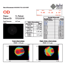 Asclepion Patient 1 |
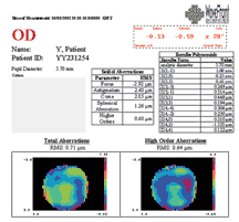 Asclepion Patient 2 |
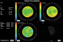 Tracey Patient 1 |
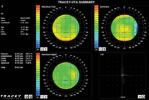 Tracey Patient 2 |
One patient was measured live at the event while the other was premeasured. Patient 1 had a best spectacle-corrected visual acuity of 20/30 and was 20/20 with contact lens correction. Patient 2 had a best spectacle-corrected visual acuity of 20/25 and was 20/20 with contact lens correction. During their presentation, the surgeons also highlighted special features of systems they use.
The session began with an overview of wavefront technologies and wavefront-guided laser surgery by Raymond Applegate, OD, PhD, professor and Borish endowed chair of optometry at the University of Houston’s College of Optometry. “Wavefront techniques have the power to explain why patients can’t see well, by revealing more information about aberrations than just sphere and cylinder,” he said.
The systemsStephen F. Brint, MD, clinical professor of ophthalmology at Tulane University School of Medicine, New Orleans, demonstrated the Alcon LadarWave CustomCornea Wavefront System. Brint described how the system works and said that it is the only system to include a registration feature. “This provides a precise locking on, so that the wavefront pattern is precisely matched to the cornea and we have a precise custom ablation. The Alcon unit and the Wavelight unit were the only two to correctly identify the live patient’s high secondary astigmatism.”
Daniel S. Durrie, MD, associate clinical professor, University of Kansas, Kansas City, demonstrated the Bausch & Lomb Zywave System’s most recent technology. “The live measurements recorded on the patient showed there was a small amount of aberration in sphere and cylinder.
There was also a tremendous amount of coma,” he said. The point-spread function was also revealing. “The point-spread function helps patients because that is what they see when they see the light, including halos and glare.”
Durrie highlighted some elements of the software, which enables doctors to adjust the spherical aberrations to see how the aberration alters the patient’s vision.
Ming X. Wang, MD, PhD, Department of Ophthalmology, Vanderbilt University, Nashville, Tennessee, demonstrated the LaserSight AstraMax Custom Corneal Topography Platform. According to Dr. Wang, new-generation corneal topography raw data are more comprehensive than with all other corneal topography systems. The corneal topography measurements consist of 35,000 data points in 0.2 seconds, he explained.
“A three-camera stereo topography versus conventional single-camera systems offers improved accuracy in anterior central and pericentral measurement of visual quality,” he said. “The pericentral measurement is the principal [measurement] … that affects visual quality.”
The system also has a two-dimensional measurement vs. a traditional one-dimensional system. “This offers improved resolution in the measurement and a more accurate anterior elevation in custom treatment,” he said.
Wang said that the No. 1 bottleneck in the marginal success of today’s custom treatment is the inaccuracy of topographic measurement. “If we are diagnosing and managing a postcorneal procedure complication, diagnosis [and treatment] should be cornea-based rather than wavefront, because that is the structure we messed up in the first place,” he said.
Howard V. Gimbel, MD, Gimbel Eye Centre, Calgary, Alberta, Canada, discussed and demonstrated the Nidek OPD-Scan, saying that the system has a time-based aberrometer rather than a positioned-based one.
The OPD (optical path difference) system has noncoherent infrared light that enters the eye and outgoing light received by photo detectors; the time difference is converted to a power map. The line of sight is coaxially oriented to the optical axis of the OPD. It covers 1,440 meridian points, he explained. “The OPD system is wavefront plus, because it does topography, autorefraction, and keratometry, which are all acquired simultaneously,” Gimbel said.
Gimbel said that in a number of difficult cases, the system has provided information to tackle highly aberrated corneas and has allowed physicians to give significant symptomatic visual improvement as well as higher-order aberration reduction.
Jeffery J. Machat, MD, national medical director, TLC Laser Eye Centers, Toronto, presented the Tracey Technologies Tracey Visual Function Analyzer. “The Tracey ray-tracing visual function analyzer is the only wavefront unit that reads 100% of complex eyes,” he said. “The system has rapid sequential ray tracing for refractive and wavefront analysis. It does this in 40 to 60 ms†. The system has the potential to do sectional and asymmetrical patterns and point-spread function assessments. The unit scans from a 2.5-mm to 8-mm optical zone. If we are going to be able to re-treat patients, we have to be able to measure irregular eyes, and with experience we can get better.”
Douglas D. Koch, MD, professor and Allen, Mosbacher, and Law Chair in Ophthalmology, Cullen Eye Institute, Baylor College of Medicine, Houston, demonstrated the Visx WaveScan Wavefront System, which uses the Hartmann-Shack aberrometer. Koch explained that in U.S. clinical trials, dilation of 6-mm pupils was in a darkened room. “It is a key feature. It is difficult if the room is not dark enough.”
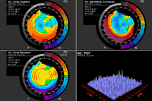 LaserSight Patient 1 |
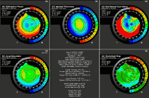 LaserSight Patient 1 |
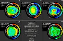 LaserSight Patient 2 |
He pointed out several of the system’s features, including the Zernike terms display and the point-spread function. “You can show this to the patient as a representation of what they might be seeing if they saw a point of light and then show them what they might see if they were corrected,” Koch said.
“We cut a PreVue lens, which the patient will later try out. This is to ensure the customized ablation will correct him as we anticipate. This was what was used in the U.S. Food and Drug Administration trials, and more than 95%-plus patients who had this done saw 20/20 +3 or better with a PreVue lens, which reassured us that we were in fact going to provide the right treatment for them. The data from our studies clearly confirm that,” he said.
Theo Seiler, MD, PhD, professor and chairman at the University Eye Clinic, in Dresden, Germany, demonstrated the WaveLight Laser Technologies Allegretto Wave Analyzer. “This is a Tscherning system, not a Hartmann-Shack sensor as the other systems are,” he said. “The system and laser have an automatic eye-tracking device to help center the measurement.”
Seiler said that, in his practice, guidelines state that if the wavefront error is less than 0.2 µm, wavefront-guided treatment or re-treatment is not done.
Robert J. Mitchell, MD, assistant clinical professor, University of Calgary, discussed and demonstrated the Asclepion wavefront aberration-supported corneal ablation (WASCA). “In my practice, we use wavefront and topographically supported customized ablation routinely. I am so convinced about the value of wavefront that we use it routinely on all patients. We do have about a dozen patients where we have used wavefront therapeutically to great benefit to the patient,” he said.
Mitchell pointed out that the system has a zonal reconstruction feature that enables it to look at the very fine orders of optical aberrations and compare them with those systems that don’t offer zonal reconstruction.
Stulting concluded the session saying, “It is clear that we have come a long way toward our understanding and ability to handle, manage, and treat this problem.”
Contact Information
Applegate: 713-743-1957, fax 713-743-2053
Brint: 504-888-2020, fax 504-888-2929
Durrie: 913-491-3737, fax 913-491-9650
Gimbel: 403-286-3022, fax 403-286-2943
Koch: 713-798-6443, fax 713-798-3027
Machat: 248-360-7601, fax 248-360-7602
Mitchell: 403-258-1773, fax 403-258-2704
Seiler: +49351-458-3381, fax +49351-458-4335
Stulting: 404-250-9700, fax 404-250-9006
Wang: 615-321-8881, fax 615-321-8874
Our new texbooks
A 501c(3) charity that has helped patients from over 40 states in the US and 55 countries, with all sight restoration surgeries performed free-of-charge.


